A Taxonomic Survey of Deep Learning Architectures for Biomedical Image Classification
Keywords:
Biomedical image classification, Deep learning, Vision transformer, Federated learning, Explainable AI, Self-supervised learningAbstract
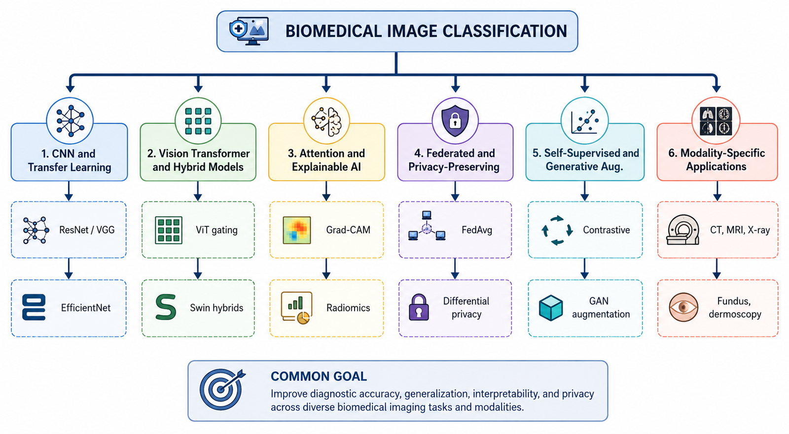
Deep learning has become central to biomedical image classification, yet the literature spans convolutional networks, vision transformers, attention-based explainability methods, federated and privacy-preserving training, self-supervised and generative-augmentation strategies, and a wide range of clinical imaging modalities, making it difficult to see the field as a coherent whole. This paper presents a taxonomic literature review of deep learning approaches for biomedical image classification published between 2022 and 2026, organizing the field into six branches: convolutional and transfer-learning architectures, vision transformer and hybrid models, attention and explainable artificial intelligence, federated and privacy-preserving learning, self-supervised and generative-augmentation strategies, and modality-specific clinical applications spanning computed tomography, magnetic resonance imaging, histopathology, dermoscopy, and retinal fundus photography. For each branch, representative works are compared in dedicated tables covering method, dataset, and reported outcome, and four architecture diagrams illustrate the taxonomy and representative pipelines. A dedicated comparison of public benchmark datasets, including MedMNIST, HAM10000, the Brain Tumor Segmentation challenge data, and ChestX-ray14, highlights how scale and annotation depth vary across modalities. The synthesis shows that architectural accuracy gains have plateaued relative to progress on privacy-preserving collaboration, label-efficient training, and trustworthy explanation, now prerequisites for clinical translation. Open challenges in cross-institution generalization, edge deployment efficiency, class imbalance, and the accuracy-interpretability trade-off are outlined to guide future research.
References
[1] J. Plested, M. Phiri, and T. Gedeon, “Deep transfer learning for image classification: a survey,” Artificial Intelligence Review, vol. 59, no. 3, 2026, doi: 10.1007/s10462-026-11491-z.
[2] N. Ajay, H. S. Mohan, I. S. Rajesh, and S. Gupta, “A deep learning and blockchain-based security framework for cloud data protection,” Journal of Discrete Mathematical Sciences and Cryptography, vol. 29, no. 6, pp. 2553-2587, 2026, doi: 10.47974/JDMSC-2823.
[3] C. Patrício, J. C. Neves, and L. F. Teixeira, “Explainable deep learning methods in medical image classification: a survey,” ACM Computing Surveys, vol. 56, no. 4, pp. 1-41, 2023, doi: 10.1145/3625287.
[4] T. R. Kulkarni and N. Dushyanth, “Early and noninvasive screening of common cardiovascular related diseases such as diabetes and cerebral infarction using photoplethysmograph signals,” Results in Optics, vol. 3, 2021, doi: 10.1016/j.rio.2021.100062.
[5] M. P. Kulkarni and M. T. Kulkarni, “Secure health monitoring of soldiers with tracking system using IoT: a survey,” International Journal of Trend in Scientific Research and Development, vol. 3, no. 4, 2019.
[6] K. R. Shwetha and K. G. Namitha, “Deep learning-based lung nodule classification and lung cancer diagnosis: a comprehensive literature review,” International Journal of Computational Intelligence in Engineering, vol. 1, no. 3, pp. 51-57, 2026.
[7] R. Wardhani and N. Nafi’iyah, “Modification CNN transfer learning for classification MRI brain tumor,” Telematika, vol. 16, no. 2, pp. 103-112, 2023, doi: 10.35671/telematika.v16i2.2272.
[8] W. Bukaita and V. Vadde, “Comparative evaluation of CNN and ResNet18 architectures for MRI-based brain tumor classification using deep learning,” Medical Research Archives, vol. 13, no. 12, 2025, doi: 10.18103/mra.v13i12.7100.
[9] W. A. Nugroho, C. Supriyanto, Pujiono, and G. F. Shidik, “Optimizing deep learning models with custom ReLU for breast cancer histopathology image classification,” Scientific Journal of Informatics, vol. 11, no. 3, pp. 849-862, 2024, doi: 10.15294/sji.v11i3.12722.
[10] U. N. Ranjitha and M. A. Gowtham, “Hybrid model using K-means clustering for volumetric quantification of lung tumor: a case study,” in Machine Learning and Autonomous Systems: Proceedings of ICMLAS 2021, Singapore: Springer Nature Singapore, 2022, pp. 527-536.
[11] G. G. Rajput, B. M. Reshmi, and I. S. Rajesh, “Automatic detection and grading of diabetic maculopathy using fundus images,” Procedia Computer Science, vol. 167, pp. 57-66, 2020, doi: 10.1016/j.procs.2020.03.182.
[12] C. T. Chandrakala, M. A. Goutham, and U. N. Ranjitha, “Statistical validation of Z-score intensity normalization in multi-modal brain MRI,” in Proceedings of the 2026 International Conference on Emerging Research in Smart Electronics and Machine Informatics, 2026, pp. 1-7.
[13] B. Dash, U. N. Ranjitha, and U. A. Kumar, “Genetic algorithm-driven hyperparameter optimization for precision sickle cell disease prognosis,” in EAI International Conference on Advanced Technologies in Electronics, Communications and Signal Processing, Cham: Springer Nature Switzerland, 2024, pp. 20-31.
[14] N. S. Shaik, T. K. Cherukuri, N. Veeranjaneulu, and J. D. Bodapati, “Medtransnet: advanced gating transformer network for medical image classification,” Machine Vision and Applications, vol. 35, no. 4, 2024, doi: 10.1007/s00138-024-01542-2.
[15] M. Kaliappan, E. Mariappan, V. Manimaran, and B. Revathi, “Unified vision transformer for multimodal medical image classification,” Biomedical Signal Processing and Control, vol. 122, 2026, doi: 10.1016/j.bspc.2026.110431.
[16] V. M. Firos, P. J. A. Alphonse, U. Fiore, and G. R. Gangadharan, “RLTNT: an explainable residual learning-based transformer model for kidney disease classification,” Image and Vision Computing, vol. 163, 2025, doi: 10.1016/j.imavis.2025.105781.
[17] R. Firdaus and A. Smerat, “XHSA-DCNet: an explainable hybrid Swin transformer and attention-guided dense convolution network for automated leukemia detection and classification,” International Journal of Computational Intelligence in Engineering, vol. 1, no. 1, pp. 43-52, 2026.
[18] Suhas S, U. N. Ranjitha, Bhuvan C U, Mahantesh K B, and Kalyan D S, “Leveraging ensemble deep learning for enhanced brain tumor analysis: integrating YOLOv8, Mask R-CNN, and U-Net,” in Proceedings of the 2024 8th International Conference on Computational System and Information Technology for Sustainable Solutions, 2024, doi: 10.1109/csitss64042.2024.10816879.
[19] J. S. Inchal, “Deep feature fusion and ensemble voting based brain tumor classification framework,” International Journal of Computational Intelligence in Engineering, vol. 1, no. 3, pp. 42-50, 2026.
[20] S. Mahawar, “Recent advances in heart disease prediction from ECG signals: a survey of machine learning, deep learning, and explainable AI approaches,” International Journal of Computational Intelligence in Engineering, vol. 1, no. 2, pp. 42-48, 2026.
[21] M. Ennab and H. Mcheick, “AI interpretability in medical imaging analysis,” Machine Learning and Knowledge Extraction, vol. 7, no. 1, p. 12, 2025, doi: 10.3390/make7010012.
[22] R. Gharaibeh, “Brain tumor detection interpretability,” Applied Computer Science, vol. 21, no. 3, pp. 182-197, 2025, doi: 10.35784/acs_7375.
[23] B. S. Sonune, R. Udaykumar, S. G. Nemane, D. P. Tulaskar, M. Bhaiyya, and H. Haick, “Interpretable deep learning in dermoscopy: an XAI-driven ensemble for skin lesion diagnosis,” IEEE Access, vol. 14, pp. 62126-62142, 2026, doi: 10.1109/access.2026.3684257.
[24] V. L. Sowmya, A. Bharathi Malakreddy, S. Natarajan, N. Prathik, and I. S. Rajesh, “Spectral entropic radiomics feature extraction (SERFE): an adaptive approach for glioblastoma disease classification,” Frontiers in Artificial Intelligence, vol. 8, 2025, doi: 10.3389/frai.2025.1583079.
[25] J. Yoon and C. Lin, “Brain tumor classification in MRI,” IEEE Access, vol. 13, pp. 154172-154202, 2025, doi: 10.1109/access.2025.3603272.
[26] K. R. Radhika, H. N. Shenoy, T. R. Vinay, H. Pooja, D. Sharma, R. Priyanka, and S. Gupta, “Communication-efficient federated learning (CEFL) for CT image classification in bandwidth-constrained wireless healthcare networks,” International Journal of Drug Delivery Technology, vol. 16, no. 13S, pp. 163-172, 2026, doi: 10.25258/IJDDT.16.13S.17.
[27] S. Gupta, I. S. Rajesh, C. Singh, U. N. Ranjitha, K. Siddesha, and P. K. Sekharamantry, “A hybrid CEFL approach using gradient sparsification and quantization for medical imaging,” International Journal of Computational Intelligence in Engineering, vol. 1, no. 1, pp. 1-15, 2026.
[28] A. S. M and C. L. Chowdhary, “Multi-modal personalized federated learning with adaptive differential privacy for medical image classification,” Scientific Reports, vol. 16, no. 1, 2026, doi: 10.1038/s41598-026-49896-z.
[29] S. Wassan, Liudajun, H. Ying, H. Dongyan, and P. Fei, “Federated learning and differential privacy: machine learning and deep learning for biomedical image data classification,” DIGITAL HEALTH, vol. 11, 2025, doi: 10.1177/20552076251358531.
[30] C. Nielsen, M. Wilms, and N. D. Forkert, “A novel gradient inversion attack framework to investigate privacy vulnerabilities during retinal image-based federated learning,” Medical Image Analysis, vol. 107, 2026, doi: 10.1016/j.media.2025.103807.
[31] V. Dachepalli and G. Sreelatha, “Privacy preserved federated deep learning for disease classification of patients medical images,” Applied Soft Computing, vol. 201, 2026, doi: 10.1016/j.asoc.2026.115512.
[32] E. Gupta and V. Gupta, “Margin-aware optimized contrastive learning for enhanced self-supervised histopathological image classification,” Health Information Science and Systems, vol. 13, no. 1, 2024, doi: 10.1007/s13755-024-00316-4.
[33] F. B. Ashraf, S. M. Alam, and S. M. Sakib, “Enhancing breast cancer classification via histopathological image analysis: leveraging self-supervised contrastive learning and transfer learning,” Heliyon, vol. 10, no. 2, 2024, doi: 10.1016/j.heliyon.2024.e24094.
[34] N. S. Punn and S. Tapaswi, “Enhancing robustness against adversarial attacks in biomedical image classification through squeezed quantized self-supervised contrastive learning,” Biomedical Signal Processing and Control, vol. 120, 2026, doi: 10.1016/j.bspc.2026.110093.
[35] S. Wang and R. Mao, “PromptMed: prompt-driven semi-supervised medical image classification with class-balanced consistency and contrastive learning,” Expert Systems with Applications, vol. 310, 2026, doi: 10.1016/j.eswa.2026.131345.
[36] Z. Liu, Q. Lv, C. H. Lee, and L. Shen, “GSDA: generative adversarial network-based semi-supervised data augmentation,” Heliyon, vol. 9, no. 9, 2023, doi: 10.1016/j.heliyon.2023.e19585.
[37] U. N. Ranjitha, D. L. Shanthi, I. S. Rajesh, C. Maithri, G. Sumit, and S. Chandrappa, “Multi-modal data fusion and dual validation for lung lesion detection and optimization,” Journal of Information Systems Engineering and Management, vol. 10, no. 40s, pp. 1027-1036, 2025.
[38] V. K. Bairagi et al., “Attention-based deep learning framework for lung nodule classification in CT images,” Symmetry, vol. 18, no. 3, p. 431, 2026, doi: 10.3390/sym18030431.
[39] T. R. Kulkarni, Bharathi, Gururaj, Aditi, and Jaiswal, “Comparison of implementation in blood cancer causes and diseases,” International Journal of Trend in Scientific Research and Development, vol. 9, no. 1, pp. 503-512, 2025.
[40] Y. M. Dalal, K. Amuthabala, and U. N. Ranjitha, “Precision medicine in nephrology: an exploratory study of machine learning models for accurate kidney disease prognosis,” in 2024 Second International Conference on Advances in Information Technology, vol. 1, 2024, pp. 1-6.
[41] I. S. Rajesh, B. M. Reshmi, A. Bharathi Malakreddy, and S. Bilakeri, “Automatic detection of hard exudate in color retinal fundus image and diabetic maculopathy grading,” IEEE Access, vol. 13, pp. 6618-6631, 2025, doi: 10.1109/access.2025.3525499.
[42] I. S. Rajesh, M. A. Bharathi, and B. M. Reshmi, “A comprehensive review on automatic diagnosis of diabetic maculopathy in retinal fundus images,” in Advances in Signal Processing and Intelligent Recognition Systems, Singapore: Springer, 2019, pp. 410-420, doi: 10.1007/978-981-13-5758-9_35.
[43] N. Kallel and A. Echtioui, “Retinal fundus image classification for diabetic retinopathy using transfer learning technique,” Signal, Image and Video Processing, vol. 18, no. 2, pp. 1143-1153, 2023, doi: 10.1007/s11760-023-02820-8.
[44] A. Shazuli and R. Saravanan, “Manta ray foraging optimizer with deep learning-based fundus image retrieval and classification for diabetic retinopathy grading,” Engineering, Technology & Applied Science Research, vol. 13, no. 5, pp. 11661-11666, 2023, doi: 10.48084/etasr.6226.
[45] G. Ramesh and S. Sathiamoorthy, “A deep learning grading classification of diabetic retinopathy on retinal fundus images with bio-inspired optimization,” Engineering, Technology & Applied Science Research, vol. 13, no. 4, pp. 11248-11252, 2023, doi: 10.48084/etasr.6033.
[46] K. S. Rashmi and A. Marndi, “Explainable deep learning ensemble model for breast cancer classification in histopathology images,” Biomedical Signal Processing and Control, vol. 123, 2026, doi: 10.1016/j.bspc.2026.110505.
[47] D. Mandair, J. S. Reis-Filho, and A. Ashworth, “Biological insights and novel biomarker discovery through deep learning approaches in breast cancer histopathology,” npj Breast Cancer, vol. 9, no. 1, 2023, doi: 10.1038/s41523-023-00518-1.
[48] A. Das and M. N. Mohanty, “Wavelet-convolutional neural network: an improved deep learning model for breast cancer detection from histopathology images,” Archives of Breast Cancer, vol. 12, no. 1, pp. 75-86, 2025, doi: 10.32768/abc.202512173-84.
[49] E. B. Kablan and S. Ayas, “Skin lesion classification from dermoscopy images using ensemble learning of ConvNeXt models,” Signal, Image and Video Processing, vol. 18, no. 8-9, pp. 6353-6361, 2024, doi: 10.1007/s11760-024-03321-y.
[50] A. K. Nugroho, R. Wardoyo, M. E. Wibowo, and H. Soebono, “Image dermoscopy skin lesion classification using deep learning method: systematic literature review,” Bulletin of Electrical Engineering and Informatics, vol. 13, no. 2, pp. 1042-1049, 2024, doi: 10.11591/eei.v13i2.6077.
[51] K. R. Radhika, S. Gupta, M. J. Oli, D. Sharma, H. Vinutha, and H. N. Shenoy, “Pancreatic tumor segmentation using conditional diffusion and graph regularization on abdominal computed tomography images,” Journal of Intelligent Decision Making and Information Science, vol. 3, no. 1s, pp. 1123-1143, 2026, doi: 10.59543/jidmis.v3.489.
[52] J. Yang, R. Shi, D. Wei, Z. Liu, L. Zhao, B. Ke, H. Pfister, and B. Ni, “MedMNIST v2: a large-scale lightweight benchmark for 2D and 3D biomedical image classification,” Scientific Data, vol. 10, no. 1, 2023, doi: 10.1038/s41597-022-01721-8.
[53] U. Lee, S. Hwang, J. Choi, and J. Choi, “Comparative analysis of active learning methods in biomedical image classification with MedMNIST benchmark,” Heliyon, vol. 12, no. 8, 2026, doi: 10.1016/j.heliyon.2026.e44917.
[54] M. Prvan, J. Musić, D. Čoko, and A. Kristić, “Lightweight neural network ensemble models for medical image classification with MedMNIST dataset,” Electronics, vol. 15, no. 7, p. 1470, 2026, doi: 10.3390/electronics15071470.
Downloads
Published
How to Cite
Issue
Section
License
Copyright (c) 2026 International Journal of Smart Technologies and Innovations

This work is licensed under a Creative Commons Attribution-NonCommercial-ShareAlike 4.0 International License.
License Terms
The International Journal of Smart Technologies and Innovations (IJSTI) publishes articles under an open-access publishing model to promote the free and global dissemination of scholarly research.
Licensing Policy
Authors publishing in IJSTI agree that:
-
The submitted work is original and does not infringe upon the copyright or intellectual property rights of others.
-
Authors retain copyright of their published articles while granting the journal the non-exclusive right to publish, archive, reproduce, and distribute the work in electronic and print formats.
-
Published articles may be accessed, downloaded, copied, shared, cited, and used for academic, educational, and non-commercial research purposes, provided proper acknowledgment and citation of the original publication are given.
-
Any commercial use, reproduction, or distribution of published content without prior written permission from the journal and/or author is prohibited.
-
Authors are responsible for obtaining permission for any copyrighted materials used in their manuscripts, including images, figures, tables, or third-party content.
-
The journal reserves the right to preserve, archive, index, and distribute published content through scholarly databases, repositories, and digital preservation systems.
-
By submitting a manuscript, authors confirm that all co-authors have approved the submission and agree to the journal’s licensing and publication policies.
Open Access Statement
IJSTI provides immediate open access to its content based on the principle that making research freely available to the public supports greater global knowledge exchange and academic advancement.
Journal Website:
IJSTI Official Website


